
We use cookies to understand how you use our site and to improve the overall user experience. This includes personalizing content and advertising. Read our Privacy Policy
Accept Cookies
T cells are vital lymphocytes in the immune system, just like the elite troops of the body's defense. It mainly develops and matures in the thymus, and then enters the blood circulation and lymphatic system, always ready to perform immune tasks. T cell receptor (TCR) is a specific recognition structure on the surface of T cells, which can be called "radar" of T cells. TCR is mainly composed of α and β chains, and each chain contains variable region and constant region. The amino acid sequence of the variable region is highly diversified, which gives TCR the ability to recognize multiple antigens.
TCR is a structure that can specifically recognize and bind antigen peptide-MHC molecular complex on the surface of T cells, and plays a core role in cellular immunity. It consists of two different polypeptide chains, usually α chain and β chain, and in a few cases γ chain and δ chain. These chains are connected by disulfide bonds, and their variable regions form unique antigen binding sites. When T cells encounter antigen peptide-MHC complex processed and presented by antigen presenting cells, TCR will accurately recognize the complex and trigger a series of intracellular signal transduction events. The specific combination of TCR and antigen peptide-MHC complex can start the activation, proliferation and differentiation of T cells, promote T cells to play a cytotoxic role and assist other immune cells, so as to eliminate cells infected by pathogens and tumor cells in the body and maintain the immune balance and health of the body.
TCR diversity is a key feature of the immune system, which is very important for the body to defend against pathogen infection and maintain immune balance. Its diversity mainly comes from the process of gene rearrangement. In the early stage of T cell development, many variable (V), diverse (D) and linked (J) gene fragments were randomly combined, and were precisely cut and linked by recombinase, resulting in a large number of different TCR coding sequences. For example, only the combination of α-chain V and J gene fragments and β-chain V, D and J gene fragments can theoretically produce more than 10 15 different TCRs. In addition, non-template addition and deletion during gene rearrangement further increase the diversity of TCR. This high diversity enables the immune system to recognize almost endless epitopes, so as to effectively deal with the invasion of various pathogens, whether known or emerging viruses, bacteria and so on. It is like a universal lock with countless keys, which ensures that it can accurately open the door to defend against various pathogens.
TCR sequencing can accurately determine the nucleotide sequence of TCR gene and reveal the diversity and specificity of T cell receptors by high-throughput sequencing analysis of TCR gene in T cells. Its principle is mainly based on the second generation sequencing (NGS) and other technologies. First, RNA or DNA from T cells is extracted, and then TCR gene fragments are enriched by reverse transcription, PCR amplification and other steps, and then these fragments are sequenced on a large scale by using the sequencing platform, thus obtaining a large amount of TCR sequence information.
Take the Next Step: Explore Related Services
Learn More
TCR Sequencing: Unraveling the Process, Applications, and Advantages
TCR is a specific antigen recognition receptor on the surface of T cells. It is usually a heterodimer composed of α and β peptide chains (a few of which are γ and δ chains) connected by disulfide bonds. Each chain contains a variable region (V region) and a constant region (C region), and the V region has a hypervariable region (complementarity determining region, CDR), which can specifically recognize antigen peptides presented by major histocompatibility complex (MHC) molecules and recognize antigens in T cells.
Overall composition of TCR
αβ TCR:95 % of T cells is composed of α chain and β chain, which belongs to heterodimer. The two chains are connected by disulfide bonds, and together with the constant CD3 molecule, they form a T cell receptor complex.
γδ TCR:5 % of T cells is composed of γ chain and δ chain, which is also a heterodimer and forms a complex with CD3 molecule.
Structure of single subunit
CDR region: The variable region of each subunit contains three highly variable complementarity determining regions (CDRs), namely CDR1, CDR2 and CDR3. CDR3 is the region mainly responsible for recognizing processed antigens. CDR1 of α subunit interacts with N-terminal of antigenic peptide, CDR1 of β subunit interacts with C-terminal of antigenic peptide, and CDR2 is considered to be mainly responsible for recognizing MHC molecules.
HV4 region: β chain has an additional hypervariable region HV4, which usually does not participate in the recognition of normal antigens, but can interact with superantigens.
Constant region: Located near the cell membrane, it contains short connecting sequences, in which cysteine residues form disulfide bonds, which are used to connect α chain and β chain or γ chain and δ chain. The constant region connects the transmembrane region and the intracellular short tail.
Transmembrane region: TCR is fixed on the cell membrane of T cells, so that TCR can receive the antigen signal outside the cell and transmit it to the cell.
Intracellular degeneracy: The intracellular region of TCR is very short, and usually does not directly participate in signal transmission, but interacts with helper molecules such as CD3, and starts intracellular signal transmission through the tyrosine activation motif (ITAM) of the immune receptor in the intracellular segment of CD3.
 Structure of a classical TCR (Gao et al., 2013)
TCR complex structure
T cell receptor complex is a transmembrane octamer, which consists of TCR dimer, CD3 δ/ε dimer, CD3 γ/ε dimer and CD247 ζ/ζ or ζ/η dimer. Each dimer is linked by the interaction between ionized amino acid residues, and the whole complex can efficiently transmit the signal received by the receptor to the cell.
TCR Classification and Features
TCR is a specific receptor for T cells to recognize antigens. TCR can be classified into α β type and γ δ type based on their different structural components and functional characteristics, and each type of TCR plays a different role in the immune system.
TCRαβ
Composition: Heterodimer composed of α chain and β chain connected by disulfide bond. Each chain contains a variable region (V region), a constant region (C region), a transmembrane region and a cytoplasmic region. Among them, V region is responsible for recognizing antigen peptide-MHC complex, and different T cells have different sequences of V region of TCR, which gives T cells the ability to recognize multiple antigens.
Expression cells: Mainly expressed on the surface of CD4+ helper T cells and CD8+cytotoxic T cells.
High specificity: TCRαβ can specifically recognize the antigen peptide presented by the major histocompatibility complex (MHC) molecules on the surface of antigen presenting cells (APC), which has high specificity and affinity and is an important basis for antigen-specific recognition in adaptive immune response.
Diversity: TCRαβ can produce extremely rich diversity through gene rearrangement and other mechanisms, and can recognize almost unlimited kinds of antigens, so that the body can cope with a variety of pathogen infections.
Role in immune response: In the process of immune response, TCRαβ can activate T cells and start a series of immune responses, such as helper T cells assisting B cells to produce antibodies and activating cytotoxic T cells to kill target cells, which plays a central role in adaptive immunity.
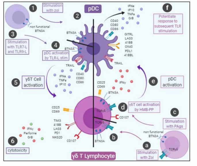 Cellular interactions between pDCs and γδ T cell (Girard et al., 2020)
TCRγδ
Composition: Heterodimers composed of γ chain and δ chain also have V region, C region, transmembrane region and cytoplasmic region. Its V region structure is also diverse, and it can recognize different antigens.
Expression cells: Mainly expressed on the surface of some special T cell subsets, such as epithelial lymphocytes in mucosal tissues such as skin and intestine.
The way to recognize antigens is unique: TCRγδ recognizes antigens independently of MHC molecules, and can directly recognize some pathogen-related molecular patterns (PAMPs) or damage-related molecular patterns (DAMPs), as well as some specific antigens on the surface of tumor cells, which plays an important role in innate immunity and early immune defense.
TCRγδ+ T cells have the functional characteristics similar to those of innate immune cells, such as fast activation, rapid production of cytokines and chemokines, participation in inflammatory response and immune regulation, rapid response in the early stage of infection or tissue injury, and initiation of immune defense.
Role of TCRγδ+ T cells in tissue immune surveillance: TCR γ δ+T cells can be used as the first line of defense to monitor and respond to local pathogen infection and abnormal cell changes, which is helpful to maintain the immune homeostasis of tissues and is of great significance in resisting mucosal infection and tumor surveillance.
Usually, each T cell only expresses one type of TCR(α β or γ δ), but there may be multiple TCR molecules on the surface of a T cell (for example, there may be about 10,000 TCR molecules on the surface of a typical T cell), and the roles of α β TCR and γ δ TCR in the immune system are different. α β TCR is mainly involved in classical antigen recognition and immune response, while γ δ TCR plays an important role in some special environments, such as infection and tumor microenvironment.
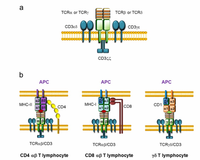 TCR structure and APC interaction (Rodriguez-Caparrós et al., 2020)
TCR gene is composed of multiple gene fragments, which are rearranged by somatic cells to form highly diversified TCR. This diversity enables TCR to recognize multiple antigens and play an important role in immune response.
TRA gene: α chain encoding TCR, located on human chromosome 14q11.2. It consists of multiple gene fragments, including variable region (V), connecting region (J) and constant region (C). During the development of T cells, these gene fragments are combined by gene rearrangement mechanism to form TCRα chain with unique specificity.
TRB gene: β chain encoding TCR, located on human chromosome 7q35. TRB gene also contains V, D (diversity region), J and C gene fragments, and its gene rearrangement process is more complicated. Through the combination of different V, D and J gene fragments, a large number of TCRβ chains with different specificities are produced.
TRG gene: γ chain encoding TCR, located on human chromosome 7p14-p15. TRG gene is similar to TRA gene in structure, and consists of V, J and C gene fragments. During the development of γδ T cells, the gene rearrangement gives γδ TCR unique antigen recognition specificity.
TRD gene: δ chain encoding TCR, located in TRA locus on chromosome 14. TRD gene contains V, D, J and C gene fragments, and its gene rearrangement is similar to TRB gene. During the development of γδ T cells, TRD gene and TRG gene are rearranged, which together determine the antigen recognition characteristics of γδ TCR.
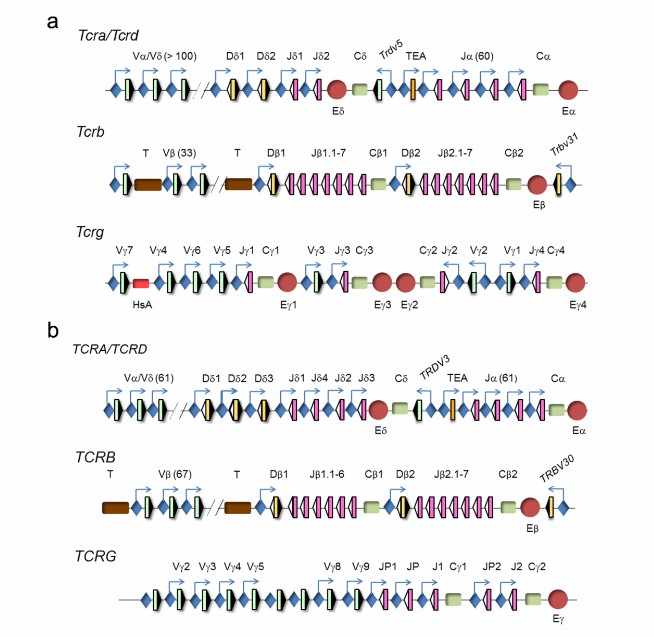 Genomic representation of mouse and human TCR loci (Rodriguez-Caparrós et al., 2020)
TCR is a specific receptor on the surface of T cells and plays a key role in the immune process. TCR can recognize the antigen peptide-MHC complex processed and presented by antigen presenting cells, and this recognition process is the initial step of T cell activation. Subsequently, T cells are activated, proliferated and differentiated into effector T cells. For example, cytotoxic T cells can directly kill cells infected by pathogens or tumor cells, while helper T cells regulate the functions of other immune cells (such as B cells, macrophages, etc.) by secreting cytokines, so as to coordinate innate and adaptive immune responses, jointly resist the invasion of pathogens and maintain the immune balance of the body. At the same time, TCR is also involved in recognizing its own antigens and preventing the immune system from attacking itself in the formation of immune tolerance.
Initiate immune response: When TCR recognizes the antigen peptide-MHC complex, T cells are activated, and begin to proliferate and differentiate, forming effector T cells and memory T cells. Effector T cells can quickly exert immune effect, while memory T cells can quickly start the second immune response when they encounter the same antigen again, resulting in a stronger and faster immune response.
Cellular immunity: CD8+ cytotoxic T cells (CTL) are the main effector cells of cellular immunity. After TCR recognizes the antigen peptide-MHC I class I complex on the surface of target cells, it can release cytotoxic substances such as perforin and granzyme, so as to make the target cells apoptosis, thus eliminating virus-infected cells and tumor cells.
Auxiliary immunity: After TCR of CD4+ helper T cells (Th cells) recognizes the antigen peptide-MHC II complex, it can help B cells to produce antibodies, enhance the phagocytosis and killing function of macrophages, and promote the activation and proliferation of CD8+ T cells by secreting cytokines, such as interleukin-2 (IL-2) and interferon-γ (IFN-γ).
TCR sequencing has important applications in many fields. In basic immunology research, it is helpful to deeply understand the development and differentiation of T cells and the formation mechanism of immune tolerance. In disease research, it can be used to analyze the immune characteristics of T cells in patients with tumors, infectious diseases and autoimmune diseases.
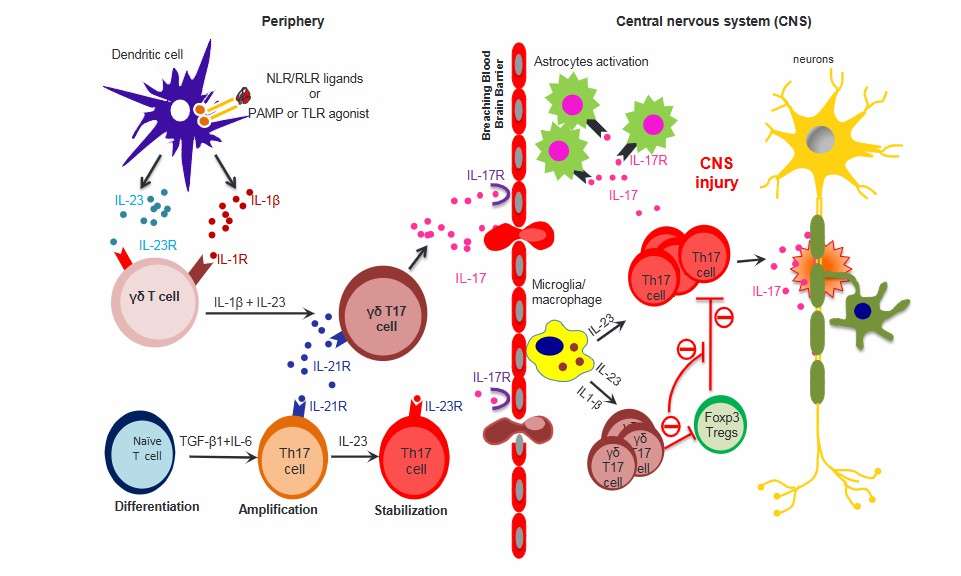 Peripherally primed γδ T cells execute their effector functions in the CNS (Malik et al., 2016)
Learn More
TCR is a characteristic mark on the surface of T lymphocytes. Heterodimer composed of two different peptide chains (α, β or γ, δ chains) connected by disulfide bonds can specifically recognize antigenic peptides presented by major histocompatibility complex (MHC) molecules, and initiate immune response processes such as activation, proliferation and differentiation of T cells. It plays a key role in cellular immunity and immune monitoring functions such as pathogen infection, tumor cell recognition and clearance.
TCR sequencing and HLA typing are two important technical means in the field of immunity, and there is a close relationship between them. In the immune reaction, the results of TCR sequencing and HLA typing complement each other. TCR sequencing can tell us the characteristics of T cells involved in immune response in the body, and HLA typing can help us to know which HLA molecules present the antigenic peptides recognized by these T cells. By combining the information of the two, we can fully understand the mechanism of immune response, which is of great significance for understanding the occurrence and development of diseases and finding therapeutic targets.
References
CD Genomics is transforming biomedical potential into precision insights through seamless sequencing and advanced bioinformatics.


