
We use cookies to understand how you use our site and to improve the overall user experience. This includes personalizing content and advertising. Read our Privacy Policy
Accept Cookies
B cells are an important kind of lymphocytes in the immune system, which play a core role in humoral immune response. B cells originated from hematopoietic stem cells in bone marrow and matured in bone marrow. Mature B cells can circulate in blood and lymphoid tissues and recognize antigens through specific receptors on the surface. When stimulated by antigen, B cells can be activated, proliferated and differentiated into plasma cells and memory B cells. Plasma cells can secrete a large number of antibodies, which can specifically bind to antigens, play a role in neutralizing toxins and regulating phagocytosis, thus eliminating pathogens. Memory B cells can be activated quickly when they encounter the same antigen again, and start a stronger and faster immune response.
B cell receptor (BCR) is a kind of mIg, which is a characteristic molecule on the surface of B cells. BCR is composed of smlg and Igα/Igβ heterodimers, and smlg is responsible for specific recognition of antigens. Its variable regions are highly diverse and can recognize various epitopes. Igα/Igβ mainly plays the role of signal transduction. When BCR binds to antigen, it will trigger a series of signal transduction events, activate the relevant signal pathways in B cells, and promote the activation, proliferation and differentiation of B cells. B cell receptor is not only a key structure for B cells to recognize antigens, but also an important basis for B cells to play their immune functions.
BCR consists of two immunoglobulin heavy chains (Igα and Igβ) and two light chains (mIg), forming a heterodimer complex. Among them, mIg is the key part of antigen recognition, which can be divided into two types: IgM and IgD. Immature B cells express mIgM, while mature B cells express both mIgM and mIgD. mIgD plays an important role in the survival and function of B cells.
mIg is the main component of BCR, which is composed of two heavy chains and two light chains connected by dimer. Heavy chain can be divided into variable region (V region), constant region (C region), transmembrane region and cytoplasmic region. The light chain has only V region and C region. Among them, V region is composed of two domains, VH and VL, and each domain has three complementary determining regions, namely CDR1, CDR2 and CDR3. All three CDRs participate in the recognition of antigens and jointly determine the antigen specificity of BCR.
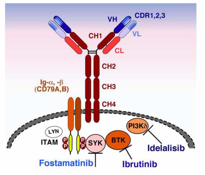
Lgα (CD79a) and Igβ (CD79b) exist as dimers, and the two peptide chains can be divided into extracellular region, transmembrane region and cytoplasmic region. Its cytoplasmic region is particularly long, with 61 amino acid residues in Igα and 48 amino acid residues in Igβ, each with an immune receptor tyrosine activation motif (ITAM), which is necessary for signal transduction and is mainly responsible for transmitting antigen-binding signals to the inside of cells.
The heavy chain and light chain are arranged in a Y-shape, and the two arms constitute the antigen binding site, which is composed of the variable regions of the heavy chain and light chain and is responsible for specifically recognizing and binding antigens. This Y-shaped structure enables BCR to bind two identical epitopes at the same time, which enhances the stability of binding.
BCR sequencing is an important technical means for in-depth study of BCR. It mainly aims at sequencing and analyzing BCR on the surface of B cells. BCR are composed of heavy chains and light chains, and their variable regions are highly diverse. By BCR sequencing, the nucleotide sequences of these variable regions can be accurately determined. The principle of this technology is based on processing the genome of a B cell population or a single B cell, enriching BCR-related gene fragments by PCR amplification, and then performing large-scale sequencing with the help of high-throughput sequencing platform.
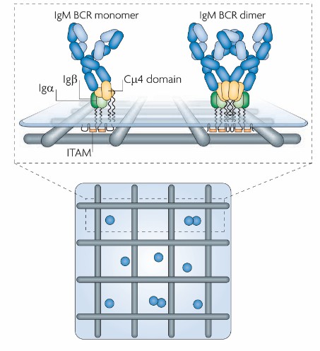 The structural organization of the BCR (Pierce et al., 2010)
Main functions
Recognition and binding of antigens: The antigen binding sites of BCR are highly diverse, and it can recognize various epitopes, including intact and natural protein antigens, polysaccharides or lipid antigens. The BCR on the surface of each B cell can only recognize a specific epitope. When the BCR binds to the corresponding antigen, the activation process of B cells is started.
Mediated antigen internalization and processing presentation: after binding with antigen, BCR internalizes antigen into cells through receptor-mediated endocytosis, forming endosomes. In endosome, antigen is degraded into small peptide fragments, and then combined with major histocompatibility complex class II molecules (MHC II) to form antigen-MHC II complex, which is transported to the cell surface and presented to helper T cells (Th cells), thus activating Th cells and assisting B cells to further proliferate, differentiate and produce antibodies.
Maintaining the survival and development of B cells: During the development of B cells, the expression and signal transmission of BCR play a key role in the survival, proliferation and differentiation of B cells. MIgM is expressed on the surface of immature B cells, and BCR signal can maintain the survival of B cells and promote their continued development and maturity if they do not encounter their own antigens. If autoantigen is recognized, negative selection will be triggered, which will lead to B cell apoptosis, thus eliminating self-reactive B cells and maintaining the self-tolerance of the immune system.
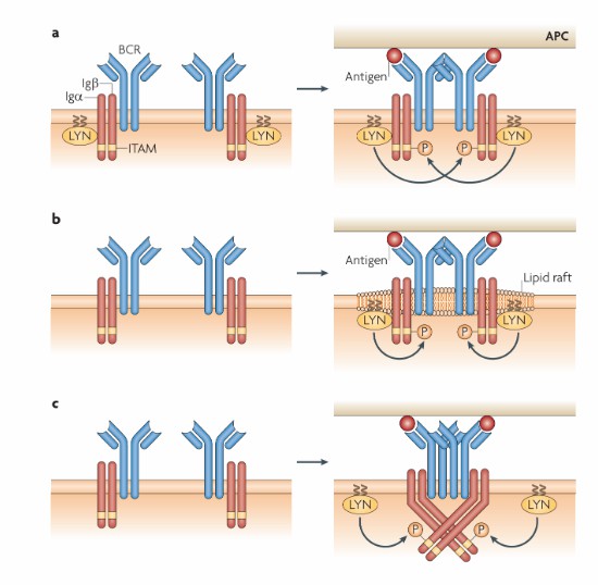 Models for how B cell receptor oligomers trigger signalling (Pierce et al., 2010)
A. Classification based on heavy chain constant region
a) IgM BCR: This is the earliest type of BCR expressed in the development of B cells. It usually exists on the surface of B cells in the form of pentamer, which has high affinity and can effectively bind antigens. IgM BCR plays an important role in the early stage of individual development and immune response, and is the first line of defense against pathogens. For example, in the early stage of infection, B cells can quickly identify and bind pathogens through IgM BCR, and start an immune response.
b) IgD-type BCR: Co-expressed with IgM-type BCR on the surface of mature B cells, similar in structure to IgM, but relatively unique in function. Although the specific function of IgD BCR is not yet fully understood, it may play a regulatory role in the activation, proliferation and differentiation of B cells, which is related to the fine recognition of antigens and the fine-tuning of immune response of B cells.
c) IgG-type BCR: It mainly exists on the surface of B cells with immune response again. IgG-type BCR has high affinity maturity and can recognize and bind antigens more accurately. It plays an important role in mediating antibody-dependent cell-mediated cytotoxicity (ADCC) and regulating phagocytosis, and is an important force in anti-infection and immune defense.
d) IgA BCR: It mainly exists on the surface of B cells in mucosa-associated lymphoid tissue and is the main effector molecule of mucosal immunity. IgA BCR can recognize and bind antigens on mucosal surface, and excrete antigens through secretory IgA, which plays a key role in resisting pathogen infection in respiratory tract, digestive tract and other mucosal parts.
e) IgE-type BCR: Under normal circumstances, the number of B cells expressing IgE-type BCR is small, but its expression will increase significantly in allergic reactions and parasitic infections. IgE-type BCR can bind to FcεRⅠ on the surface of mast cells and basophils, so that these cells are sensitized. When the same antigen enters the body again and binds to specific IgE on the surface of sensitized target cells, these cells can be promoted to synthesize and release bioactive substances, causing type I hypersensitivity.
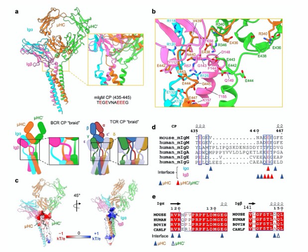 IgM BCR assembly at the CP regions (Dong et al., 2022)
B. Classification according to antigen recognition specificity
a) Multi-reactive BCR: This kind of BCR can recognize many different epitopes and has relatively broad specificity. Multi-reactive BCR plays a certain role in innate immunity, which can quickly identify and combine multiple pathogen-related molecular patterns (PAMPs) or damage-related molecular patterns (DAMPs) to start innate immune response and provide early signals for the start of adaptive immune response.
b) Monospecific BCR: It only has a highly specific recognition ability for a specific antigen epitope. In the adaptive immune response, B cells of monospecific BCR will undergo clonal proliferation and differentiation after encountering the corresponding antigen, producing a large number of specific antibodies against the antigen, thus achieving accurate elimination of specific pathogens.
Take the Next Step: Explore Related Services
Learn More
BCR-Seq: A Comprehensive Overview of Process, Advantages, Challenges, and Applications
BCR signal transduction is a key process for B cells to recognize antigens and start immune response. The following is a detailed introduction.
Antigen recognition and BCR cross-linking: When B cells encounter antigens that are complementary to their mIg specificity, mIg specifically binds to the antigen. If the antigen is multivalent, it can combine with multiple BCRs at the same time, so that BCRs can be crosslinked. This is the initial step of BCR signal transduction.
Activation of tyrosine kinases: After BCR cross-linking, tyrosine kinases Lyn, Fyn and Blk in the cytoplasm were recruited near the BCR complex, and the immune receptor tyrosine activation motif (ITAM) on the intracellular segments of Igα and Igβ was phosphorylated. Phosphorylated ITAM recruits and activates another tyrosine kinase Syk, which further phosphorylates downstream signal molecules, thus starting the signal transduction cascade.
Activation of phospholipase C-γ (PLC-γ): The activated Syk phosphorylates adapter proteins BCAP and SLP-65, which recruit and activate PLC-γ. PLC-γ hydrolyzed phosphatidylinositol-4,5-diphosphate (PIP2) into diacylglycerol (DAG) and inositol triphosphate (IP3).
Activation of calcium signaling pathway: IP3 binds to IP3 receptor on endoplasmic reticulum, which releases calcium ion (Ca), leading to the increase of intracellular Ca concentration. Ca²⁺ binds to calmodulin, activates calcineurin, and then dephosphorizes nuclear factor-activated T cells (NFAT) and enters the nucleus, regulating the transcription of related genes.
Activation of protein kinase C (PKC): DAG recruits and activates PKC on plasma membrane. PKC activates transcription factors such as nuclear factor κB (NF-κB) by phosphorylating a series of substrates, and promotes the expression of genes related to B cell activation, proliferation and differentiation.
Activation of MAPK signal pathway: BCR signal can also activate mitogen-activated protein kinase (MAPK) signal pathway by activating small G protein Ras, including extracellular signal-regulated kinase (ERK), c-Jun amino-terminal kinase (JNK) and p38 MAPK. These MAPK enter the nucleus, phosphorylate transcription factors and regulate cell proliferation, differentiation and apoptosis.
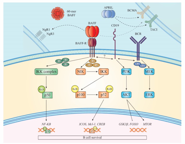 BCR signaling pathways (Damianidou et al., 2022)
BCR diversity is the basis for the body to recognize and respond to a large number of different antigens, and its mechanism mainly includes the following aspects.
Gene rearrangement: During the development of B cells, the variable region (V), diversity region (D) and junction region (J) of heavy chain gene (IGH) will be rearranged. V, D and J gene fragments each have multiple copies, and they will be randomly combined during rearrangement. For example, a V gene fragment can be combined with different D and J gene fragments, resulting in many different heavy chain variable region sequences. Light chain genes are divided into κ chain and λ chain genes, which are rearranged in a similar way to heavy chain, and are also variable regions of light chain composed of V and J gene fragments. There are also many choices for V and J gene fragments of light chain genes, and the diversity of BCR is further increased by random combination.
Linkage diversity: In the process of heavy chain gene rearrangement, terminal deoxynucleotidyl transferase (TdT) can randomly add some non-template-encoded nucleotides at the junction when V, D and J gene fragments are connected, which is called N-region insertion. These additional inserted nucleotides further increase the diversity of BCR heavy chain variable regions. In the process of gene rearrangement, after DNA double strand breaks, some nucleotides may be deleted or added in the process of repair, which is called nucleotide addition in P region. This change will also lead to the diversity of BCR sequences.
High-frequency somatic mutation: B cells enter the germinal center after being stimulated by antigen, and the variable region of BCR gene will undergo high-frequency somatic mutation. These mutations can change the binding affinity between BCR and antigen, so that B cells can produce high affinity BCR which is more suitable for antigen, further increase the diversity of BCR, and contribute to the fine recognition and immune response of different antigens.
Combination diversity: heavy chain gene rearrangement and light chain gene rearrangement are independent processes, and many different heavy chains and light chains produced after heavy chain and light chain gene rearrangement can be randomly combined to form a large number of different BCR. BCR is usually composed of mIg and Igα/Igβ, and different combinations of mIg and Igα/Igβ can also produce certain diversity.
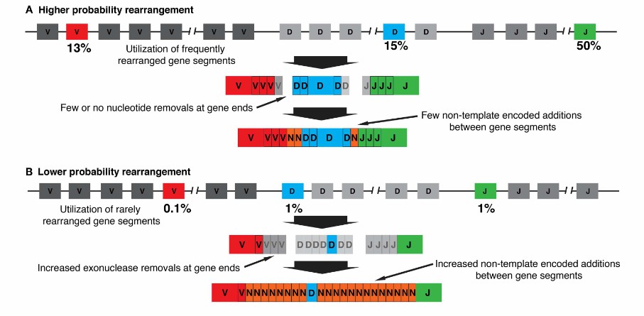 The probabilities of generating particular V(D)J rearrangements (Jackson et al., 2013)
Targeted therapeutic targets: Targeted drugs targeting BCR and its signaling pathway have become an important means to treat a variety of B-cell malignant tumors. Rituximab, for example, can specifically bind CD20 (a part of BCR complex) on the surface of B cells and kill tumor cells through various mechanisms, which can be used to treat diseases such as non-Hodgkin's lymphoma.
Immunotherapy regulation: By regulating BCR signal, immune response can be enhanced or inhibited, which can be used to treat immune-related diseases. For example, in the treatment of rheumatoid arthritis, we can try to suppress BCR signal, reduce the production of autoantibodies and alleviate inflammatory reaction.
CAR-T cell therapy: In CAR-T cell therapy technology, BCR-related molecules such as CD19 are often targeted, and patients' autologous T cells are separated and extracted, and genetically engineered to express CAR targeting CD19. After being transfused into patients, B-cell tumor cells expressing CD19 can be specifically recognized and killed.
In basic immunology research, BCR sequencing is helpful to deeply understand the development, differentiation and immune response mechanism of B cells and reveal how B cells recognize and respond to various antigens. In the aspect of disease diagnosis and treatment, it can be used for the diagnosis, typing and condition monitoring of hematological diseases such as lymphoma, and it can also provide key information for the development of treatment methods based on B cell immunity.
 Mechanism of BCR activation in chronic lymphocytic leukemia (Burger et al., 2013)
BCR is a membrane-bound immunoglobulin on the surface of B lymphocytes, which is the key structure for B cells to recognize antigens. It is composed of smlg (mIg) and Igα/Igβ (CD79a/CD79b) heterodimers. mIg is responsible for the specific binding of antigen, while Igα/Igβ is responsible for transmitting the signal generated by antigen binding. There are many types of BCR, which are mainly distinguished according to the categories of smlg, such as IgM and IgD. When BCR binds to antigen, it can activate the signal pathway in B cells, and promote the activation, proliferation and differentiation of B cells into plasma cells and memory B cells. Plasma cells secrete antibodies to play a humoral immune function, and memory B cells quickly start a stronger immune response when they encounter the same antigen again. BCR plays a key role in recognizing and eliminating pathogens and maintaining immune balance.
BCR sequencing is closely related to tumor microenvironment profiling in many aspects. The analysis of tumor microenvironment needs the combination of many technologies. As one of them, BCR sequencing can be combined with transcriptome sequencing and protein omics analysis to provide more comprehensive data for the study of tumor microenvironment.
References
CD Genomics is transforming biomedical potential into precision insights through seamless sequencing and advanced bioinformatics.


