
 Sample Submission Guidelines
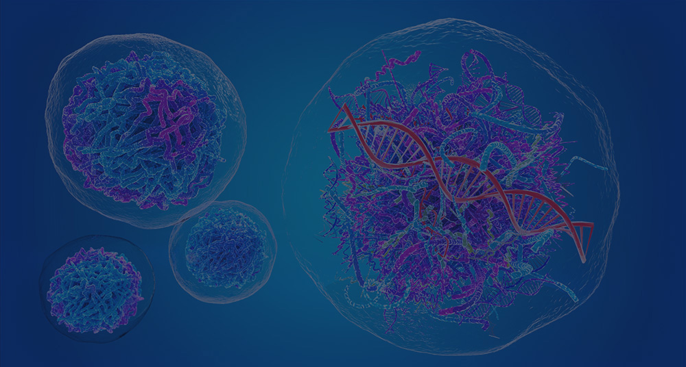
RNA virus belongs to the first class of virus classification, and its core feature is that the genetic material is RNA rather than DNA. This kind of virus widely exists in nature, including many pathogens that have great influence on human health, such as influenza virus, HIV, novel coronavirus (SARS-CoV-2) and so on. According to the types of nucleic acid strands, RNA viruses can be divided into single-stranded RNA (ssRNA) and double-stranded RNA (dsRNA), among which single-stranded RNA is further divided into positive strand (+ssRNA) and negative strand (-ssRNA).
What is Viral RNA
RNA virus is a kind of virus with ribonucleic acid (RNA) as its genetic material, which occupies a unique position in the field of biological inheritance and evolution. Unlike organisms that take deoxyribonucleic acid (DNA) as their genetic material, the genetic information of RNA viruses is carried on RNA molecules. They can’t transmit genetic information through semiconservative replication of DNA like cell biology, but rely on special mechanisms such as RNA replication or reverse transcription. After infecting the host cell, RNA virus uses various conditions of the host cell, such as enzyme system and ribosome, to replicate its own genetic material and synthesize protein, thus completing the process of virus proliferation.
RNA virus is composed of protein capsid and RNA genetic material inside, which has no cell structure and belongs to non-cell microorganism. Protein capsid are composed of repeated protein subunits, which play a role in protecting nucleic acids and mediating host cell invasion.
Nucleic acid: The nucleic acid of RNA virus can be single-stranded or double-stranded. ssRNA viruses can be divided into positive-stranded and negative-stranded viruses. The genomic RNA of +ssRNA virus itself has the function of mRNA, which can be directly recognized by ribosomes in host cells and translated into protein. The genome RNA of -ssRNA virus needs to be transcribed into complementary positive-strand RNA before translation. dsRNA virus needs to rely on its own RNA-dependent RNA polymerase, and after the virus enters the host cell, it uses one strand of double-stranded RNA as a template to transcribe mRNA.
Protein capsid: Also known as capsid, it is composed of multiple protein subunits. Its main function is to protect the nucleic acid of the virus from external factors such as nucleases, and to help the virus recognize and combine with the host cell. Different RNA viruses have different capsids in shape and structure, and some are spirally symmetric, such as tobacco mosaic virus. Some are icosahedral, such as poliovirus.

The overall shape of the CCMV capsid viewed from outside (Zhang et al., 2004)
Envelope: Some RNA viruses have envelope structure besides capsid. Envelope is a lipid bilayer membrane derived from host cell membrane or nuclear membrane, which is embedded with glycoprotein encoded by virus. Envelope can help the virus escape the recognition of the host immune system, and it is also closely related to the infectivity of the virus.
Next-generation sequencing (NGS) technologies, such as RNA-Seq, can be used to analyze the viral genome, identify mutations, and monitor changes in gene expression during infection. In addition, Sanger sequencing is often used for targeted sequencing of specific regions of the viral genome, providing a more detailed view of mutations and genetic variations in small-scale studies or confirmatory analyses.
The Main Types of Viral RNA
The main types of viral RNA can be classified and analyzed from many angles.
+ssRNA virus
The genome of +ssRNA viruses is directly translated as mRNA without reverse transcription. These viruses include enterovirus, rhinovirus, cardiovirus, aphthous virus and hepatovirus. The 3′ untranslated region (UTR) of +ssRNA viruses usually has important regulatory functions, such as the formation of stem loops, pseudoknots and TLS, which play an important role in the translation and replication of viruses.
-ssRNA virus
The genome of -ssRNA virus needs to be transcribed into +ssRNA through reverse transcription to be translated. Such viruses include coronavirus (such as SARS-CoV-2), measles virus and mumps virus. The genome of -ssRNA virus usually contains a complete 5′ cap structure and 3′ polyadenylation tail, which play a key role in the transcription and replication of the virus.
dsRNA virus
The genome of dsRNA virus consists of two complementary RNA chains, which usually exist in circular form. Such viruses include plant viruses and some animal viruses. For example, papaya ringspot virus (PRSV) is a typical double-stranded RNA virus.
Retrovirus
The genome of retrovirus is ssRNA, but its replication needs to transcribe RNA into DNA by reverse transcriptase, and then integrate it into the genome of host cell. Such as human immunodeficiency virus (HIV) and mouse retrovirus.

Classification of RNA viruses is shown with different colors (Khan et al., 2021)
How Does Viral RNA Replicate
The replication of viral RNA is a complex and diverse process, and the replication mechanism is different according to the type of viral RNA (positive or negative strand) and its genome structure.
Replication of +ssRNA virus
The genome of +ssRNA virus can be directly translated as mRNA, so its replication process is relatively simple. +ssRNA viruses usually use ribosomes and transcription mechanisms of host cells for direct translation and replication.
- Replication mechanism: The genome of +ssRNA virus can be directly translated into protein as mRNA after entering the host cell. These protein include RNA-dependent RNA polymerase (RdRp), which is responsible for the synthesis of complementary negative-strand RNA. The -ssRNA is then used as a template to synthesize new +ssRNA molecules, thus completing the replication process.
- Replication site: the replication of +ssRNA virus usually occurs in cytoplasm, using the ribosome and transcription mechanism of host cells.
- Replication intermediates: In some cases, +ssRNA viruses will form dsRNA intermediates (RI), which will be used as templates for further RNA synthesis.

Process of +ssRNA virus replication (Kang et al., 2018)
Replication of -ssRNA virus
-ssRNA virus needs to transcribe genomic RNA into +ssRNA first, and then use +ssRNA as a template for translation and replication.
- Transcription process: after the genome RNA of -ssRNA virus enters the host cell, it is first recognized by RdRp encoded in virus particles and begins to be transcribed. During transcription, RdRp uses -ssRNA as a template to synthesize +ssRNA.
- Translation and replication: +ssRNA is then translated into protein as mRNA, and these protein include RdRp and other necessary replication factors. These factors are assembled into replication complexes, and new -ssRNA is synthesized by using +ssRNA as a template.
- Replication site: The replication of -ssRNA virus usually occurs in cytoplasm, but some viruses (such as flavivirus) may replicate on endoplasmic reticulum membrane.
Retrovirus replication
Retroviruses (such as HIV) adopt a unique replication mechanism, which converts +ssRNA into DNA by reverse transcriptase, and then transcribes DNA into RNA.
- Reverse transcription process: Reverse transcriptase first reverse-transcribes +ssRNA into DNA, forming dsDNA intermediate. Subsequently, dsDNA intermediate is integrated into the genome of the host cell, and mRNA is produced through the transcription mechanism of the host cell.
- Replication site: The reverse transcription process takes place in cytoplasm, and after DNA is integrated into the genome of the host cell, it is replicated through the transcription mechanism of the host cell.
The role of replication complex
Replication Complex plays a key role in the replication process of both positive-strand and negative-strand RNA viruses. Replication complex usually consists of protein encoded by virus (such as RdRp, protease, etc.) and ribosomes and tRNA provided by host cells.
- Function: Replication complex is responsible for recognizing template RNA, synthesizing new RNA chains and assembling new virus particles.
- Protection mechanism: Some viruses (such as influenza virus) use vesicles formed by the host cell membrane as replication sites to protect the replication process from the attack of the host immune system.

RNA virus replication machineries (Tao et al., 2010)
Errors and mutations in the process of replication
Due to the lack of proofreading function of RNA-dependent RNA polymerase, errors are prone to occur in the process of viral RNA replication, resulting in high mutation rate. This feature enables viruses to quickly adapt to environmental changes and form virus populations through mutation accumulation.
Generally speaking, the replication process of RNA viruses is ingenious and complicated. They use the resources of host cells to amplify their own genetic material and produce a large number of virus particles through a unique mechanism, which also makes RNA viruses have a strong ability to spread and survive in nature.
The Principle of Viral RNA Extraction
The principle of virus RNA extraction is mainly based on the following key steps: lysis, separation, purification and so on. The purpose of these steps is to release RNA from the sample and separate it from other cellular components for subsequent detection or analysis.
- Lysis: First, the cell membrane or other structures in the sample need to be destroyed to release RNA. This is usually done by using lysis buffer, which may contain acidic or alkaline substances (such as acidic buffer or chloroform) and detergents (such as SDS or Triton X-100) to destroy cell membranes and release RNA.
- Virus protein is separated from nucleic acid: Virus consists of protein shell and internal nucleic acid (RNA or DNA). After cell splitting, virus protein needs to be separated from RNA. Protease K and other enzymes are usually added, which can degrade the protein shell of the virus and expose the virus RNA. At the same time, we can also use the solubility difference between nucleic acid and protein in different chemical environments to promote the separation. For example, in high-salt or low-salt solution, the solubility of nucleic acid and protein will be different, thus achieving preliminary separation.
- RNA binding and purification: The affinity of RNA with specific substances is used to capture and purify RNA. It is common to use silica gel membranes or magnetic beads. Silica gel membrane can specifically bind RNA under the condition of high salt and low pH value, and RNA can be eluted under the condition of low salt and high pH value. The surface of magnetic beads is usually modified with groups that can bind to RNA. Under the action of magnetic field, the magnetic beads bound with RNA can be separated from other impurities, and then the RNA is eluted from the magnetic beads by eluent to obtain purified viral RNA.

Clinical efficacy evaluation of the viral RNA extraction methods (Hongjaisee et al., 2022)
- Impurities removal: During the extraction process, impurities such as protein and genomic DNA may remain. Residual DNA can be degraded by adding RNase-free DNase, and then insoluble impurities can be removed by centrifugation, filtration and other methods, and finally high-purity viral RNA can be obtained for subsequent experiments such as detection and analysis.
Different extraction methods may be slightly different, but the overall principle is similar, and each has its own advantages and disadvantages. Choosing the appropriate method depends on the sample type, target virus type and laboratory conditions.
How is Viral RNA Extracted
There are many methods to extract viral RNA, and the specific steps and reagents used vary according to the sample type and detection purpose. The following are some common virus RNA extraction methods and their detailed steps:
TRIzol method
- Sample treatment: Cell or tissue samples are added into a centrifuge tube containing Trizol reagent, and for cell samples, 1 ml of Trizol is added to every 106 cells. For tissue samples, add 1 ml of trizol per 50-100mg of tissue. The sample is fully cracked by repeated blowing with homogenizer or pipette.
- Phase separation: After the sample is cracked, it is allowed to stand at room temperature for 5 minutes to completely dissociate the ribosome. Then add 0.2ml chloroform to every 1 ml of trizol reagent, cover the tube cover tightly, shake violently for 15 seconds, and let it stand at room temperature for 2-3 minutes. Then centrifuge at 12000g for 15 minutes at 4℃, and the sample will be divided into three layers. The upper layer is a colorless and transparent water phase containing RNA. The middle layer is a white protein layer. The lower layer is a red organic phase containing DNA and protein.
- RNA precipitation: Carefully suck the upper water phase and transfer it to a new centrifuge tube. To avoid sucking the protein in the middle layer, don’t suck the lower organic phase. Add 0.5ml isopropyl alcohol to 1ml Trizol reagent, mix it upside down, and let it stand at room temperature for 10 minutes to precipitate RNA. Then centrifuge at 12000g for 10 minutes at 4℃, and white RNA precipitation will appear at the bottom of the centrifuge tube.
- RNA washing: Discard the supernatant, add 1 ml of 75% ethanol to wash the RNA precipitate, vortex the precipitate to suspend, and then centrifuge at 7500 g at 4 ℃ for 5 minutes. Discard the supernatant, reverse the centrifuge tube on the absorbent paper, and let the ethanol evaporate naturally to dry the RNA precipitate, but be careful not to over-dry, so as not to affect the dissolution of RNA.
- RNA dissolution: Add proper amount of RNase-free water or TE buffer, gently blow or incubate in 55-60℃ water bath for 10 minutes to completely dissolve RNA.

Schematic showing the standard TRIzol RNA extraction procedures (Lee et al., 2011)
Magnetic bead method
- Sample preparation: Cell or tissue samples are crushed and lysed, and the method is similar to that of Trizol method, but special magnetic bead lysis solution is used.
- Magnetic bead binding: Add an appropriate amount of magnetic bead suspension to the lysate, and the surface of magnetic beads has groups that can specifically bind to RNA. Gently mix and incubate at a certain temperature and time to fully combine RNA with magnetic beads.
- Magnetic field separation: Put the centrifugal tube on the magnetic frame, so that the magnetic beads are adsorbed on the tube wall, and then carefully suck the supernatant to remove impurities.
- Washing magnetic beads: Wash magnetic beads for many times with washing buffer to remove unbound impurities and protein. After each washing, it is necessary to put the centrifuge tube on the magnetic frame and suck the supernatant.
- RNA elution: Add an appropriate amount of elution buffer to the magnetic beads and incubate for a period of time at an appropriate temperature to elute RNA from the magnetic beads. Then remove the centrifuge tube from the magnetic frame, mix it gently, put it back into the magnetic frame, and suck the eluent containing RNA into the new centrifuge tube.

Manufacture of esiRNA by means of a magnetic bead-integrated chip (Wang et al., 2012)
Reverse transcription
Reverse transcription is not a method of directly extracting RNA, but a method of synthesizing cDNA using the extracted RNA as a template, provided that the RNA extraction has been completed, and the following steps are taken:
- Preparation of RNA template: Take a proper amount of extracted RNA samples and add RNase-free water to adjust the volume to the required amount.
- Preparation of reaction system: RNA template, reverse transcription buffer, dNTP mixture, reverse transcriptase, primer (random primer, Oligo (dT) primer or specific primer) and RNase-free water are added in the PCR tube in turn to prepare the reaction system.
- Reverse transcription reaction: Put the PCR tube into the PCR instrument and perform reverse transcription reaction according to the set procedure. Generally, it includes incubating at 42-50℃ for 30-60 minutes for reverse transcription, then heating at 70-85℃ for 5-10 minutes to inactivate reverse transcriptase and terminate the reaction. After the reaction, the product obtained is cDNA, which can be used in subsequent experiments such as qPCR and QCPR.

Methods for detection of RNA virus (MARTÍNEZ et al., 2022)
Conclusion
RNA virus is a kind of virus with ribonucleic acid (RNA) as its genetic material, which occupies an important position in the virus family. According to the characteristics of genome, RNA viruses can be divided into single-stranded RNA viruses and double-stranded RNA viruses, and single-stranded RNA viruses are further divided into positive-stranded RNA viruses and negative-stranded RNA viruses. The genomic RNA of positive-strand RNA virus itself can be used as messenger RNA(mRNA) to directly translate and synthesize protein. The genomic RNA of negative-strand RNA virus needs to be transcribed into complementary positive-strand RNA before it can play the role of mRNA. This classification difference is very important for understanding the infection mechanism and replication process of the virus.
In the research of RNA virus, extracting virus RNA is a key step. There are various extraction methods, such as Trizol method and magnetic bead method. Although the principles and operations of these extraction methods are different, their purpose is to obtain high-purity viral RNA, which provides a basis for further study of virus gene sequence, transcription regulation, pathogenic mechanism and development of diagnostic reagents and vaccines.
References
- Khan J, Asoom LIA., et al. "Evolution of RNA viruses from SARS to SARS-CoV-2 and diagnostic techniques for COVID-19: a review." Beni Suef Univ J Basic Appl Sci. 2021 10(1):60 https://doi.org/10.1186/s43088-021-00150-7
- Zhang D, Konecny R, Baker NA, McCammon JA. "Electrostatic interaction between RNA and protein capsid in cowpea chlorotic mottle virus simulated by a coarse-grain RNA model and a Monte Carlo approach." Biopolymers. 2004 (4):325-337 https://doi.org/10.1002/bip.20120
- Kang S, Brown HM, Hwang S. "Direct Antiviral Mechanisms of Interferon-Gamma." Immune Netw. 2018 18(5):e33 https://doi.org/10.4110/in.2018.18.e33
- Tao YJ, Ye Q. "RNA Virus Replication Complexes." PLoS Pathog 2010 6 (7): e1000943 https://doi.org/10.1371/journal.ppat.1000943
- Hongjaisee S, Jabjainai Y., et al. "Comparison of Simple RNA Extraction Methods for Molecular Diagnosis of Hepatitis C Virus in Plasma." Diagnostics (Basel). 2022 12(7):1599 https://doi.org/10.3390/diagnostics12071599
- Lee, Juliana T.Y., et al. "Simple Modifications to Standard TRIzol® Protocol Allow High-Yield RNA Extraction from Cells on Resorbable Materials." Journal of Biomaterials and Nanobiotechnology (2011): 41-48 https://doi.org/10.4236/JBNB.2011.21006
- Wang Z, Yang J., et al. "A Magnetic Bead-Integrated Chip for the Large Scale Manufacture of Normalized esiRNAs." PLoS ONE 2012 7(6): e39419 https://doi.org/10.1371/journal.pone.0039419
- MARTÍNEZ GÓMEZ, Alejandro. "Plan de contingencia, estrategias post COVID-19." Medicina e Investigación Universidad Autónoma del Estado de México. (2022): 42-48 http://orcid.org/0000-0003-4677-7985